

北京大学熊汗青博士课题组
Dr. Hanqing Xiong's lab
at Peking University

北京大学熊汗青博士课题组
Dr. Hanqing Xiong's lab
at Peking University
Current Projects in Our Lab
Advanced Spectroscopy Methods for Probing the Health State of Each Individual.

Exploring new molecular spectroscopy
We are exploring next-generation universal spectroscopy methods with advanced laser technology. A good example is the transient stimulated Raman scattering (T-SRS) spectroscopy technique recently developed in our lab.
As illustrated in the left figure, two identical dual-band femtosecond pulse pairs (pump and Stokes) are used to excite vibrational transitions in molecules with a controlled time delay. The two transition amplitudes in the vibrationally excited states undergo quantum interference with the phase determined by the delay, which in turn governs the final probability of vibrational transitions. Scanning the time delay modulates this probability and ultimately encodes the free-induction decays (FIDs) of excited vibrational modes (i.e., Raman modes) into the spectroscopic readout [in this case, stimulated Raman loss (SRL)]. Fourier transform of the FIDs then yields a standard Raman spectrum with natural-linewidth-limited spectral resolution (meaning the spectral linewidths are determined solely by the vibrational coherence lifetimes).
The beauty of T-SRS lies in its unique advantages: (i) It enables shot-noise-limited SRL measurements, resulting in high sensitivity, negligible background, and spectra identical to benchmark spontaneous Raman. (ii) Its dual-band configuration allows for collinear excitation geometry, enabling high-throughput and high-resolution imaging with high-repetition-rate lasers, high-NA objectives, and relatively low pulse energy. (iii) It unifies large bandwidth with high spectral resolution without sacrificing acquisition speed (i.e., spectral bandwith is only determined by excitation laser bandwidths). These advantages make T-SRS a game-changer for universal spectroscopic measurements and hyperspectral Raman imaging.
Related publications by our group:
Nature Photonics, (2026) DOI: 10.1038/s41566-025-01841-8;
Light: Science & Applications 13.1 (2024): 70;
JACS 145.14 (2023): 7758-7762.

Profiling clinical health states with advanced spectroscopy
Diseases usually result in obvious changes in the body's metabolic state, which are reflected in the molecular compositions of biofluids (such as serum and urine). Monitoring the composition of different biofluids can therefore help decipher the health status of an individual. Raman spectroscopy is perhaps the most ideal method for probing changes in chemical composition of biofluids, as it is label-free, low-cost, and relatively fast compared to other techniques. However, conventional spontaneous Raman scattering suffers from weak signals and fluorescence interference, making it challenging for high-throughput and highly consistent biofluid profiling. In contrast, the T-SRS spectroscopy developed in our lab offers more than 100 times faster throughput than spontaneous Raman, natural-linewidth-limited spectral resolution, broad bandwidth, and state-of-the-art sensitivity, while being inherently immune to fluorescence. These features make it the ideal tool for acquiring big data on human health status.
As shown in the proof-of-concept test on the right, high-throughput T-SRS measurements (~1 s dwell time per sample) of approximately 2,300 clinical serum samples enabled systematic profiling of eleven critical biomarkers commonly used to assess liver and kidney function, cardiovascular diseases, and other conditions. This demonstrates the feasibility of generating high-quality, large-scale datasets for data-driven health science. In addition to serum profiling, rapid cholangiocarcinoma diagnosis based on T-SRS profiling of the C-H stretching band in clinical bile samples was also demonstrated to be highly efficient and accurate. As we delve deeper in this direction, we believe T-SRS is poised to serve as a "foundational tool" for generating high-quality core big data, empowering next-generation AI-driven healthcare and other application scenarios.
Related publications by our group:
Nature Photonics, (2026) DOI: 10.1038/s41566-025-01841-8;
Analytical Chemistry, 2025, 97(15): 8499-8505.

Chemical imaging in biomedical applications
As mentioned above, Raman spectroscopy is an ideal label-free contrast mechanism for deciphering the chemical composition of biological systems. However, conventional confocal spontaneous Raman microscopy is known for its low imaging speed (~1 s per pixel dwell time) and suffers from fluorescence interference. The development of T-SRS brings new opportunities for easy access to full-range Raman imaging. It boosts the imaging speed by more than two orders of magnitude while providing even higher spectral quality (i.e., background-free and better spectral resolution).
As an example, the left figure shows a typical full-range hyperspectral Raman image of mouse liver tissue acquired by T-SRS. The entire imaging session took only a few minutes, compared to hours of data acquisition required by conventional confocal spontaneous Raman microscopy.
Related publications by our group:
Nature Photonics, (2026) DOI: 10.1038/s41566-025-01841-8;
Light: Science & Applications 13.1 (2024): 70;

Advanced instruments for spectroscopy and imaging
Developing devices and instruments that best fit our spectroscopy applications has always been a central theme of our research. A few examples are listed on the left.
(i) Advanced Laser System: The femtosecond-pulsed optical parametric oscillator (OPO, panel a) designed in our lab enables free tuning of multiple laser beams across the near-infrared region (panel b).
(ii) The ultrafast Michelson interferometer for delay scanning (panel c).
(iii) The ultra-low noise resonant photodetector that enables shot-noise-limited detection with ~50 μs dwell time for laser power down to ~0.1 mW (panel d).
Related publications by our group:
Optics Express, 2026, 34(1), 598-607;
Light: Science & Applications 13.1 (2024): 70;
JACS 145.14 (2023): 7758-7762.
Our people
Arranged in chronological order of joining date (except for the lab manager).

熊汗青 Hanqing Xiong, the Principal Investigator
Hanqing's current research interest is focused on developing new methods and instruments for the large-scale profiling of the health states of individuals, using his specialized knowledge in spectral physics, optics, and engineering. He is also considering trying new topics in understanding how the brain functions in the near future.
He likes reading and daydreaming when he's not working. He's also a pretty good amateur music composer.
Email:xiong.hanqing@pku.edu.cn

余乔智 Qiaozhi Yu, PhD student (G4)
Undergraduate: Sun Yat-sen University, majoring in Physics
His research interests lie at the intersection of photonics and molecular imaging, with a focus on stimulated Raman scattering, nonlinear optics, and spectral analysis. Outside the lab, he enjoys staying active through tennis and badminton.

郭瑾 Jin Guo, PhD student (G3)
Undergraduate: Northwestern Polytechnical University, majoring in MEMS
Jin received her B.E. from the School of Mechanical and Electrical Engineering at Northwestern Polytechnical University. As a former member of the university's model aircraft team, she contributed to the solar-powered unmanned aerial vehicle (UAV) project, which cultivated her practical engineering skills in mechanical modeling, structural analysis, and system integration. Grounded in a background of semiconductor-related engineering and hands-on project experience, she is particularly interested in exploring the underlying physical principles and building advanced optical systems. Beyond academic work, she enjoys skiing, fitness training, and playing the piano.

张浩杰 Haojie Zhang, PhD student (G3)
Undergraduate: Harbin institute of technology, majoring in Communication Engineering
His research interests include fluorescence imaging, stimulated Raman scattering, and analog circuit design. In his daily life, he enjoys fitness and traveling, which provide a continuous source of motivation for his scientific research.

俞文皓 Wenhao Yu, PhD student (G2)
Undergraduate: Peking University, majoring in Biomedical Engineering
Wenhao received his B.E. from the College of Engineering at Peking University. With a background in electronics and skills in software development, he is particularly interested in tackling practical engineering problems that arise in scientific research. Beyond academic work, he enjoys skateboarding and music production, both of which continually shape his curiosity about systems, structure, and creativity.

王家凯 Jiakai Wang, PhD student (G1)
Undergraduate: Sun Yat-sen University, majoring in Biomedical Engineering
Research Interests and Expertise: Biomedical applications of time-domain stimulated Raman spectroscopy, embedded system development, and host computer software design
His hobbies: Playing badminton and billiards

苏立川 Lichuan Su, PhD student (G0, Admitted)
Undergraduate: Tianjin University, major in Measuring and Controlling Technologies and Instruments
Research Interests: Integrating electronics, optics, precision mechanisms and control theory for instrument design
Hobbies: Socializing, playing LEGO, and reading novels

许高萌 Gaomeng Xu, PhD student (G0, Admitted)
Undergraduate: Dalian University of Technology, majoring in Applied Physics.
His hobbies include singing, basketball, and billiards.

周子懿 Ziyi Zhou, Undergraduate student
Undergraduate: Peking University, majoring in Biomedical Engineering
With a passion for biomedical optics, Ziyi is currently building a white-light interferometer, gaining hands-on experience in optical instrumentation and signal analysis. Her academic interests focus on the convergence of photonics and molecular imaging, with a desire to explore stimulated Raman scattering and nonlinear optical techniques in future research.
Outside the lab, she enjoys painting, crafting, and playing various ball sports to stay creative and active.
She's always hoping to learn — feel free to reach out if you'd like to chat about optics or research!

王劭逊 Shaoxun Wang, Lab manager
Education: East China Normal University, M.S. in Software Engineering
Outside the lab, Shaoxun is a professional musician with fantastic skill on the flute (formerly ranked top 10 in a national-level competition). He is the former Executive Secretary-General of the China Flute Association.
Shaoxun is co-hired by four labs at NBIC: the labs of He Sun, Shuai Na, Shuijing Tang, and Hanqing Xiong. He is the financial manager for our lab.
Email: wsx@pku.edu.cn
Alumni
- 庄晨洁 Chenjie Zhuang, Peking University, majoring in Biomedical Engineering, 2022-2024. She conducted undergraduate research and completed her thesis in our lab. She is now a PhD student at Columbia University.
Students Transferred or Quit
Hanqing's suggestion: PhD students who try but find that their interests are not aligned with the lab are encouraged to transfer to another lab. However, transfer does not always solve the problem, since the opportunities for moving to a well-suited lab are usually not that high. Transfer and quitting waste considerable time and energy for both the student and the PI, as well as lab resources and opportunities, and are best avoided at the beginning of enrollment. Therefore, it is highly recommended that students gain a full understanding of the lab before enrolling. Dr. Hanqing Xiong provides strong support for visiting students' projects, especially for potential PhD candidates.
Dr. Hanqing Xiong 熊汗青 博士
Diving into science and technology for a calm mind.

Assistant Professor, National Biomedical Imaging Center, College of Future Technology, Peking University
Dr. Hanqing Xiong received his Bachelor's degree from the Department of Biomedical Engineering at Huazhong University of Science and Technology in 2012, followed by his Master's training at the Wuhan National Laboratory for Optoelectronics, Huazhong University of Science and Technology until 2015. He then pursued his doctoral studies at Columbia University, where he trained as a spectroscopic physicist and earned his Ph.D. from the Department of Chemistry in 2020. He joined PKU as an independent researcher in 2021. His research focuses on ultrafast molecular vibrational spectroscopy and its applications in biomedical detection and imaging. He has published systematic research innovations in the fields of (i) coherent Raman spectroscopy & imaging and (ii) mesoscale connectomics.
He enjoys doing experiments, learning, and teaching, and spends most of his time on them (leaving little time and energy for socializing). Luckily, he was awarded by the National Young Talent Project (QB) of China in 2023, and was selected to receive long-term stable support from 2026. These awards enable him to freely explore the science he likes over the next few years.
Dr. Hanqing Xiong now spends half the week at the Huairou campus and half the week at the main campus. His office addresses:
Room 409, Building 2, National Biomedical Imaging Science Center (Huairou Campus)
339E, Yingjie Exchange Center (Main Campus)
Email: xiong.hanqing@pku.edu.cn
Daily Life in the Lab
When diving into the present, every day is so great!
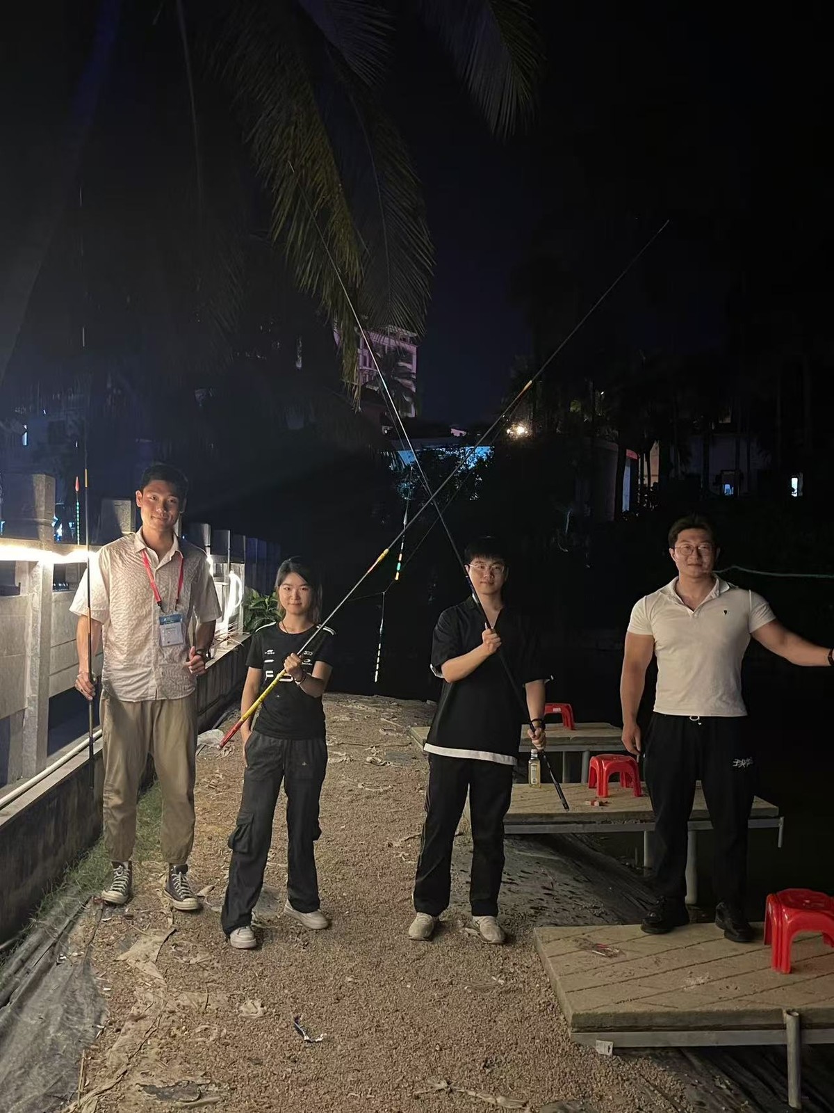
Qiaozhi, Jin, Jiakai, and Haojie (from left to right) went fishing in Sanya during the PIBM2026 Conference. June 19, 2026.
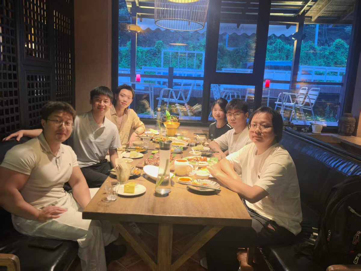
A nice dinner for group members at the Huairou Campus. June 1, 2026.
From left to right: Haojie, Lichuan, Gaomeng, Jin, Jiakai, and Hanqing.

Blooming of peonies on the main campus of PKU. April 18, 2026.

New Year's blessing from Hanqing. February 18, 2026.
Every year, Hanqing rewrites a new one for the lab.

Hanqing lighted a UV pumped visable OPO on January 4, 2026.
This is the finalized version of the previous proof-of-concept prototype.

The New Year's light show at PKU on January 1, 2026.
Shot in the middle of the night.

Jin is working with the SuperB-SRS system on September 18, 2025.
She built several versions of the SuperB-SRS system over the past year until it worked well.

A nice night view outside the window of the Huairou Campus, April 22, 2025.
More buildings are under construction.

Prototype dual-OPO system built by Hanqing, October 10, 2022.
The orange light is SFG built from the OPO output.
Join us!
A good day to succeed!
Prospective PhD Students:
We invite talented undergraduates interested in our research to visit for a lab tour. If you would like to be considered, please email Hanqing directly. Admission typically takes place through the Summer Campus program. Candidates with a master's degree and a strong background in physics and engineering are also welcome to apply in the winter, interviews are held every spring semester. For administrative updates, please monitor the website of the College of Future Technology. We have 1-2 position in 2026.
Prospective Postdocs:
We are hiring candidates with expertise in ultrafast laser physics, microbiology, or artificial intelligence. We have 1-2 position in 2026.
Undergraduate students:
We encourage any student interested in our research to apply. We have multiple positions available for undergraduates.
For all the cases above: please get in touch with Hanqing ASAP (xiong.hanqing@pku.edu.cn).
Our Lab address:
Room 316, Building 2
National Biomedical Imaging Science Center, Peking University
North Dajian Road, Huairou Science City
Huaibei Town, Huairou District
Beijing, China, 101400


 京公网安备11010802047866号